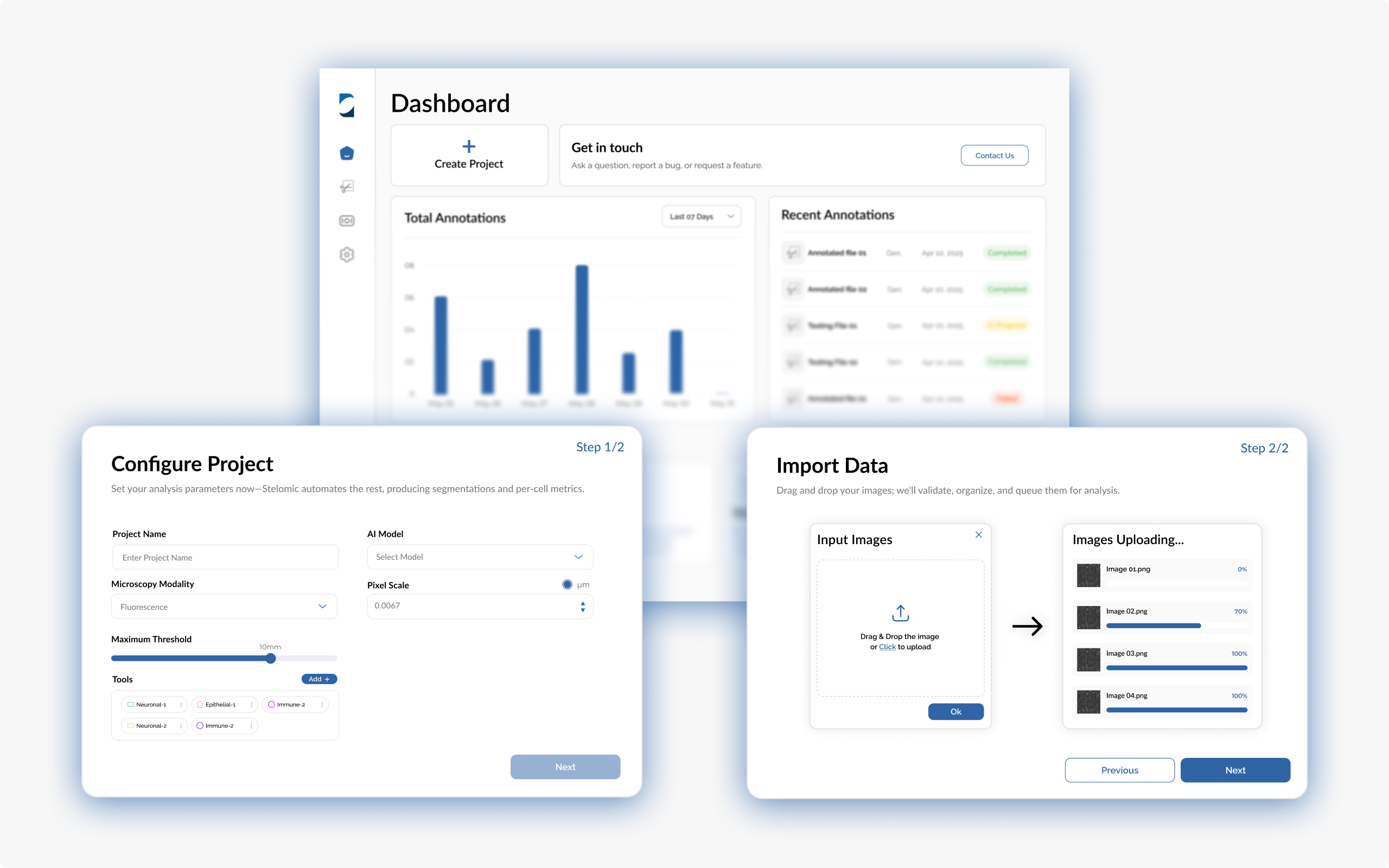
Automated cell segmentation,
from upload to analytics in minutes
Expert-trained AI labeling and quantitative metrics for microscopy imaging.
Trusted by scientists at leading research labs and biotech companies


Validated across 12 unique microscopy datasets

Spanning 40 cell types and 5 microscopy modalities

Compared to traditional annotation pipelines and methods
Manual annotation wastes hours,
outdated software is inaccurate,
and DIY solutions are hard to build.
Stelomic shifts time from labeling to discovery.

Developed by biomedical AI experts,
tuned for research-grade accuracy.

One-Click Metrics
Instantly outputs cell count, area, perimeter, centroid, circularity, confluency, and solidity. Reduces human error and speeds up analysis—offering publication-grade performance.

Ready-to-Use AI Model
Start analyzing immediately with our pre-trained AI model. Built on thousands of annotated images for high accuracy. No coding, training, or AI expertise needed.

Private & Secure
Designed to support HIPAA and GDPR compliance. Your files stay private and encrypted, never shared.

Compatibility
Supports brightfield, darkfield, phase contrast, widefield, fluorescence, confocal, and EM—validated across 40+ cell types, all on one platform.

Performance Analytics
Per-image and batch-level validation—accuracy, precision, recall, specificity, Dice/F1, IoU, and Hausdorff distance—available with custom training.
Level up microscopy image analysis with Stelomic Copilot
Discover how to turn raw files into research-ready data in minutes.