 Stelomic
Stelomic Copilot
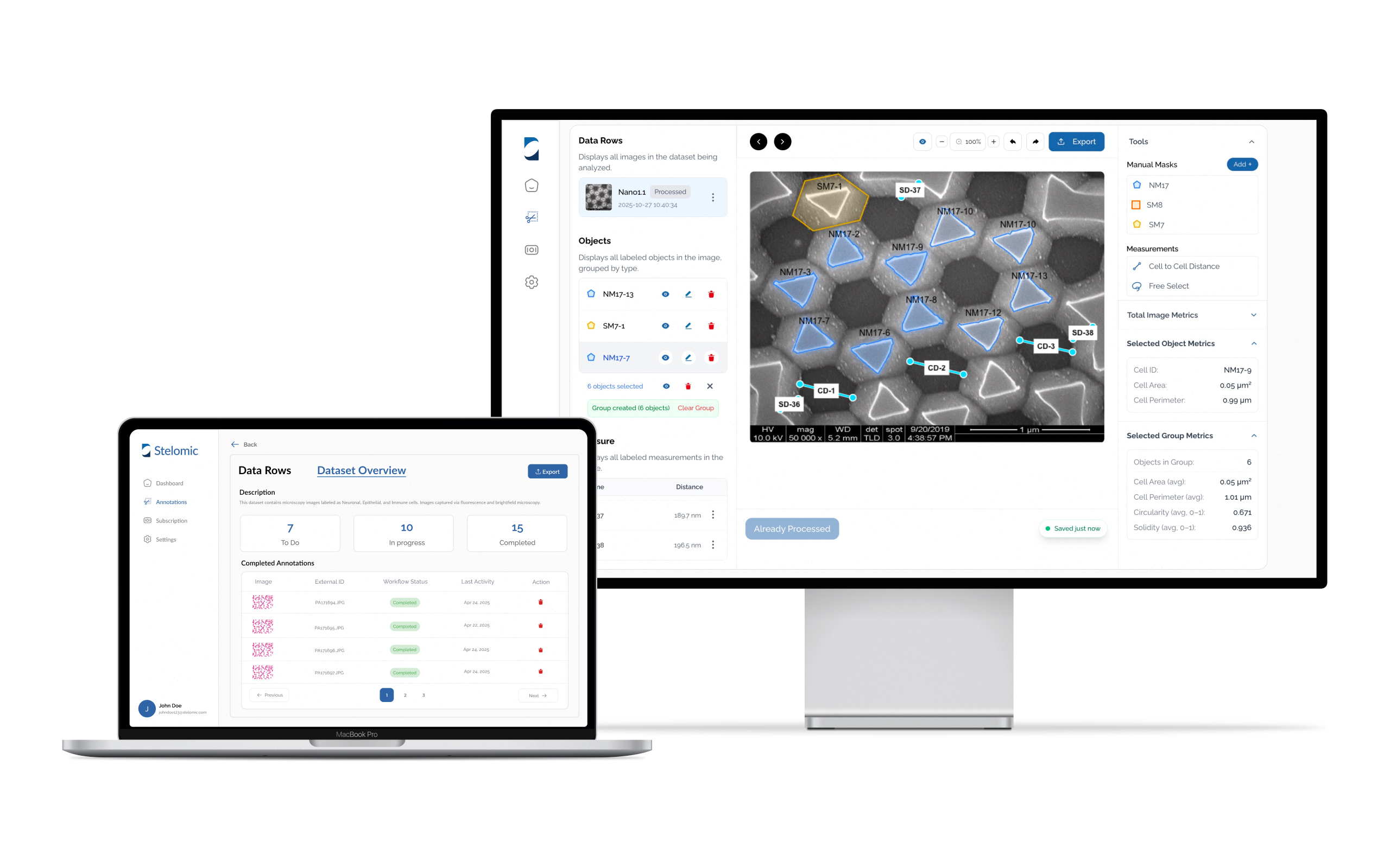
Automate your microscopy workflows with transformer-enhanced deep learning.
1
Upload
Start a project, drag-and-drop your microscopy images, and organize your files.
2
Analyze
Our AI model performs instant segmentation, generating masks and object metrics.
3
Verify
Review overlays, refine with manual edits, and add annotations.
4
Export
Download results—masks, overlays, and tables—for analysis and reporting.

98.7% Accuracy
Validated across 12 unique microscopy datasets

100K+ Cells Analyzed
Spanning 40 cell types and 5 microscopy modalities

30x Faster Processing
Compared to traditional annotation pipelines and methods
Case Studies
Real World Performance
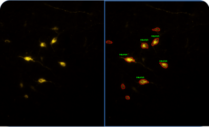
Neuronal Cell Detection
Microscopy Type: Fluorescence microscope
Model Used: Darkfield
Key Metrics:
- Accuracy: 97.8% vs manual ground truth
- Cell Count: 11
- Avg. Circularity: 0.574
- Avg. Nearest-neighbour distance: 12.4 μm

RBC & WBC Segmentation
Microscopy Type: High-magnification Light microscope
Model Used: Brightfield
Key Metrics:
- Accuracy: 98.4%
- RBC Count: 16
- WBC Count: 1
- Avg. Cell Area: 4973.56 μm²
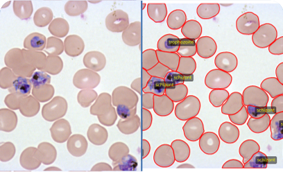
Malaria-Infected Cell Identification
Microscopy Type: Brightfield
Model Used: Generalist
Key Metrics:
- Accuracy: 96.9%
- Cell Count: 540
- Infected Cells: 18
- Avg. Solidity: 0.91
Ask us anything
We're here to simplify your workflow. Let us develop a solution for your application.